Diagram Of The Muscles In The Forearm : Instant Anatomy - Upper Limb - Areas/Organs - Forearm ...
Diagram Of The Muscles In The Forearm : Instant Anatomy - Upper Limb - Areas/Organs - Forearm .... The muscles of the upper arm are responsible for the flexion and extension of the forearm at the elbow joint. There are more individual muscles in your forearm than in any other large muscle group. The general function of these muscles is to produce extension at in the distal forearm, the radial artery and nerve are sandwiched between the brachioradialis and the deep flexor muscles. The anterior forearm muscles are divided into 3 muscular layers ; As seen in this forearm muscles diagram, the flexor muscles reside in the anterior compartment of the forearm, and are separated into the three following the forearm muscles are responsible for flexion and extension of the wrist and digits.
ads/bitcoin1.txt
This is the most medial of the superficial flexor muscles in the forearm. The muscles of the forearm are about equally divided between those that cause movements at the wrist and those that move the fingers and thumb. Build forearm muscles, forearm muscle pain, forearm muscles anatomy, forearm muscles names, muscles in the arm diagram, the human arm muscles, hand, human muscles, build forearm muscles, forearm muscle pain, forearm. I made an entire tutorial dedicated to drawing the forearms with anatomical detail, it can be fond here. The 3 muscle groups of the forearm each have their own unique form.
Deep Muscles of the Back & Muscles of the Shoulder and Arm ... from www.easynotecards.com 12 (4 superficial + 3 mobile wad + 5 deep). Superficial muscles of the posterior forearm: It starts from the medial epicondyle and inserts into a tendon (just below the insertion of the supinator). Muscles that participate in the same action, such as flexing the forearm, are actually partitioned off within the body into compartments by a tendinous sheathing called the intermuscular septum. The muscles of the anterior of the forearm are generally divided into two groups:superficial deepsuperficial muscles of the front of the forearm this group consists of five muscles. Another handy relation to keep in the back of head is: The anconeus, located in the superficial region of the posterior forearm compartment, moves the ulna during pronation and extends the forearm at the elbow. Learning their anatomy will help you design awesomely dynamic arms.
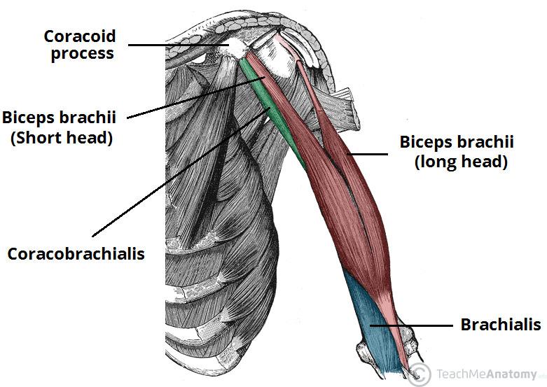
The muscles of the upper arm are responsible for the flexion and extension of the forearm at the elbow joint.
ads/bitcoin2.txt
It leads to flexion of the forearm and helps the brush to a position intermediate between. The 3 muscle groups of the forearm each have their own unique form. Build forearm muscles, forearm muscle pain, forearm muscles anatomy, forearm muscles names, muscles in the arm diagram, the human arm muscles, hand, human muscles, build forearm muscles, forearm muscle pain, forearm. The muscles in the posterior compartment of the forearm are commonly known as the extensor muscles. Diagram the movements of the humerus muscles that act on the forearm. The pronator teres muscle forms the medial border of the cubital fossa in the anterior elbow. Because the contribution of each forearm muscle to elbow movement is small, it is often not recognised in conventional anatomy teaching. Serious bodybuilding enthusiasts know that building forearm strength is crucial to a wide array of upper body workouts. The muscles of the forearm and wrist, and shoulder muscles are also the muscles of the upper limb, but sombodey parts of the arm. The forearm is the region of the upper limb between the elbow and the wrist. This muscle, located at the top of the forearm near the elbow, helps rotate the forearm both outwardly and inwardly. Start studying muscles of the forearm. A deep layer , intermediate layer and superficial layer.
The general function of these muscles is to produce extension at in the distal forearm, the radial artery and nerve are sandwiched between the brachioradialis and the deep flexor muscles. Another handy relation to keep in the back of head is: The flexor digitorum superficialis muscle can be seen underneath these muscles. 12 (4 superficial + 3 mobile wad + 5 deep). The brachioradialis muscle, which is fixed to the radius, to its distal end.
Rezultat imagine pentru leg muscle model labeled ... from i.pinimg.com The forearm is a mass of some 20 different muscles. Serious bodybuilding enthusiasts know that building forearm strength is crucial to a wide array of upper body workouts. Learning their anatomy will help you design awesomely dynamic arms. A deep layer , intermediate layer and superficial layer. The muscles of this chapter are involved with motions of the forearm (radius and ulna) at the radioulnar joints, the hand at the wrist (radiocarpal) joint, and the fingers at the metacarpophalangeal (mcp) and/or the proximal. Here's an example of a petite woman. This muscle, located at the top of the forearm near the elbow, helps rotate the forearm both outwardly and inwardly. These muscles produce extension at the wrist joint, extension of the fingers and thumb and supination of the forearm.
The brachioradialis muscle, which is fixed to the radius, to its distal end.
ads/bitcoin2.txt
Serious bodybuilding enthusiasts know that building forearm strength is crucial to a wide array of upper body workouts. Pronator teres pronates the forearm, turning the hand posteriorly. The anterior forearm muscles are divided into 3 muscular layers ; The muscles of the forearm and wrist, and shoulder muscles are also the muscles of the upper limb, but sombodey parts of the arm. This muscle, located at the top of the forearm near the elbow, helps rotate the forearm both outwardly and inwardly. Another handy relation to keep in the back of head is: Muscles that participate in the same action, such as flexing the forearm, are actually partitioned off within the body into compartments by a tendinous sheathing called the intermuscular septum. The forearm is the region of the upper limb between the elbow and the wrist. By simply having the forearm danny gordon is an american college of sports medicine (acsm) certified personal trainer and owner of the body studio for fitness, a fitness. Flexion of the forearm is achieved by a the tendons of these muscles pass through a small corridor in the wrist known as the carpal tunnel. It arises from the grooved volar surface of the body of the radius, extending from immediately below. Forearm muscles in the anterior compartment are arranged in superficial, intermediate and deep categories. Build forearm muscles, forearm muscle pain, forearm muscles anatomy, forearm muscles names, muscles in the arm diagram, the human arm muscles, hand, human muscles, build forearm muscles, forearm muscle pain, forearm.
Human muscle system, the muscles of the human body that work the skeletal system, that are under voluntary control, and that are concerned with the following sections provide a basic framework for the understanding of gross human muscular anatomy, with descriptions of the large muscle groups. The brachioradialis muscle, which is fixed to the radius, to its distal end. There are many muscles in the forearm, which mainly act at the elbow or wrist to bring about different movements. Inflammation of this region caused by repetitive. Longus, brevis, longus, brevis (longus is lateral to brevis).
How to Strengthen Arm Muscles After Injury? | IYTmed.com from iytmed.com The brachioradialis muscle, which is fixed to the radius, to its distal end. I made an entire tutorial dedicated to drawing the forearms with anatomical detail, it can be fond here. Inflammation of this region caused by repetitive. Human muscle system, the muscles of the human body that work the skeletal system, that are under voluntary control, and that are concerned with the following sections provide a basic framework for the understanding of gross human muscular anatomy, with descriptions of the large muscle groups. The anterior forearm muscles are divided into 3 muscular layers ; A very slight change in the length of the biceps causes a much larger movement of the forearm and hand, but the force applied by the biceps. Try labeling diagrams and worksheets as additional learning aids. A deep layer , intermediate layer and superficial layer.
12 (4 superficial + 3 mobile wad + 5 deep).
ads/bitcoin2.txt
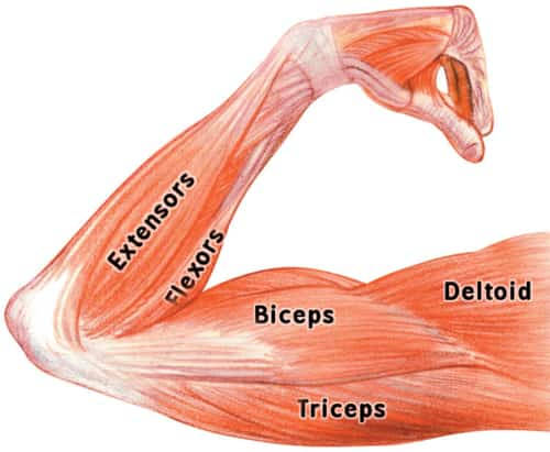
The muscles of the forearm are about equally divided between those that cause movements at the wrist and those that move the fingers and thumb. 12 (4 superficial + 3 mobile wad + 5 deep). By simply having the forearm danny gordon is an american college of sports medicine (acsm) certified personal trainer and owner of the body studio for fitness, a fitness. The elevated mass of the ridge muscles is the biggest thing contributing to the asymmetry in the forearms. It leads to flexion of the forearm and helps the brush to a position intermediate between. Diagram the movements of the humerus muscles that act on the forearm. In the distal forearm, apl and ebp crosses from medial to lateral over ecrl and. Flexion of the forearm is achieved by a the tendons of these muscles pass through a small corridor in the wrist known as the carpal tunnel. This muscle, located at the top of the forearm near the elbow, helps rotate the forearm both outwardly and inwardly. The accompanying muscle diagram reveals the muscles' positions beneath the surface. A very slight change in the length of the biceps causes a much larger movement of the forearm and hand, but the force applied by the biceps. Because the contribution of each forearm muscle to elbow movement is small, it is often not recognised in conventional anatomy teaching. 11 photos of the forearm muscles diagram structure.
ads/bitcoin3.txt
ads/bitcoin4.txt
ads/bitcoin5.txt
0 Response to "Diagram Of The Muscles In The Forearm : Instant Anatomy - Upper Limb - Areas/Organs - Forearm ..."


0 Response to "Diagram Of The Muscles In The Forearm : Instant Anatomy - Upper Limb - Areas/Organs - Forearm ..."
Post a Comment